
Mycoplasma are a member of the Mollicutes, an order within the Tenericutes phyla that do not contain a peptidoglycan cell wall. This feature is unique to Mollicutes and many of these including Mycoplasma are chemo-organotrophic organisms.

As such these bacteria are more susceptible to osmotic stress and are resistant to Beta-Lactam derived antibiotics such as Penicillin’s and cephalosporins, although they are sensitive to antibiotics which target other metabolic processes (tetracycline and erythromycin).


Cells of Mollicutes contain only a trilaminar membrane as a barrier to the outside environment, this is fortified with lipoglycans and sterols to compensate for the lack of stability caused by no cell wall. Additionally, this feature means Mollicutes exhibit pleomorphism and can exist in many forms (pear shaped, filamentous) – also allowing them to pass through biological filters.
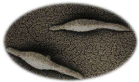
Mycoplasma colonies tend to be < 1mm in diameter displaying a typical umbonate “fried-egg” apperance – However other colonies may have cauliflower-like or smooth colony surfaces with smooth, irregular or scalloped margins.
Genera within the Mollicutes are parasitic organisms infecting a variety of eukaryotic hosts. This parasitic nature may in part be caused by their small genomes; Mollicutes have the smallest genomes of any bacteria (see diagram 2) which greatly reduces their metabolic functions, meaning they rely on hosts for specific growth requirements. Mollicutes and more specifically Mycoplasmas, can be considered as a ‘bridge’ between virus and bacteria; some viruses known as giant viruses (giruses) have genomes more than double the size of Mycoplasma genitalium (580kb).

Some clinically important genera within the Mollicutes include:
- Mycoplasma
- Ureaplasma
- Spiroplasma
Mycoplasma and Ureaplasma are parasites of humans, insects and other small animals (e.g. fish). They colonize the mucosal layers of the body including the urogenital tract. Mycoplasma are also common lab contaminants of cell lines.
Mycoplasmas are usually non-motile, however some species possess specialized structures which allow them to exhibit gliding motility, the attachment organelle of Mycoplasma pneumoniae is and example of this. Additionally, Mycoplasma mobile is the fastest Mycoplasma and can travel at 2.0 – 4.5 µm/s – or 3 -7 times its cell length per second.
Spiroplasma also exhibit motility, this time by corkscrew movement through liquid. Spiroplasma are again parasites of many organisms including plants. The motility of these organisms is mediated by contracting elements of their internal filaments – this structure responds via chemotaxis.
FURTHER READING:
An interesting note on simple organisms:
Titan, one of Saturn’s moons is the only moon within the solar system that has its own atmosphere, this comprises mainly of methane and nitrogen.
In 2012 an experiment was carried out (Horst. M.S., et al, 2012) where the atmosphere of Titan was simulated, in such experiment it was found that all 5 bases of both DNA and RNA were produced; as well as a few amino acids via various photochemical reactions in parts of the upper and lower atmosphere. If true, nucleotides could literally rain on the surface of Titan or be dissipated into space.
Also detected by NASA on titan was the presence of acrylonitrile – vinyl cyanide that can potentially form flexible structures closely resembling cell membranes. Should the nucleotides and vinyl cyanide come into contact, a very simple organism could be produced, providing basic replication strategies exist – the creation of a very rudimentary virus/bacteria.
(Hörst, S.M, et al. 2012. ‘Formation of Amino Acids and Nucleotide Bases in a Titan Atmosphere Simulation Experiment’. Astrobiology. 12(9), pp 809 – 817. https://dx.doi.org/10.1089%2Fast.2011.0623)
Image references:
Image 1: http://www.99eyao.com/english/d/mycoplasma.html
Image 2: https://en.wikipedia.org/wiki/Penicillin
Image 3: https://en.wikipedia.org/wiki/Erythromycin
https://www.ncbi.nlm.nih.gov/pubmed/8905075
https://www.ncbi.nlm.nih.gov/pmc/articles/PMC98941/
http://www.sciencedirect.com/science/book/9780126520507
https://link.springer.com/book/10.1007%2Fb113360
http://www.antimicrobe.org/m06.asp
https://www.ncbi.nlm.nih.gov/books/NBK7637/
http://www.sciencedirect.com/science/article/pii/B9780702040894000561
http://www.sciencedirect.com/science/article/pii/S0769260984800538
http://www.cell.com/current-biology/fulltext/S0960-9822(05)00684-6
http://www.cell.com/current-biology/pdf/S0960-9822(05)00684-6.pdf
https://link.springer.com/chapter/10.1007/978-1-4615-2478-6_2
https://www.ncbi.nlm.nih.gov/pmc/articles/PMC3584481/
http://www.sciencedirect.com/science/article/pii/S0960982205006846?via%3Dihub
https://www.ncbi.nlm.nih.gov/pmc/articles/PMC1303205/
http://jb.asm.org/content/197/18/2952.full
https://www.nature.com/articles/nrmicro.2016.197/figures/1
